What is the Retina?
The retina is a light-sensitive, paper-thin tissue that lines the interior back of the eye. It is attached to the optic nerve, which is tethered to the brain. Light enters the eye through the cornea, is bent, and then bent again by the lens as it is refracted and focused to a precise point on the retina.
The retina is filled with millions of photoreceptor cells called rods and cones that convert light into nerve impulses, which travel through the optic nerve to the brain and allow you to see.
The macula, the center or “bull’s eye” of the retina, has the highest concentration of cones, and cones, unlike rods which give peripheral vision, are critical for sharp, central vision. The more light that can be focused to a precise pinpoint on the macula, the better will be your vision.
Common Retinal Conditions
Although the retina is resilient, several common conditions can occur which can affect the health of the retina.
These include:
Diabetic Retinopathy
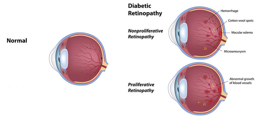
In patients with diabetes, especially with long-standing diabetes, small blood vessels called microaneurysms can form. These tiny blood vessels can leak fluid and bleed, causing vision impairment.
Proliferative diabetic retinopathy goes one step further, as a branching network of blood vessels grow out of control and eventually leak fluid and bleed.

Treatment for different stages of diabetic retinopathy is more successful than ever and can include laser treatment, intraocular injections, and surgery to remove any blood interfering with vision. Good control of blood sugar and A1C, as well as regular eye checkups, can reduce the likelihood of vision loss.
Retinal Detachment
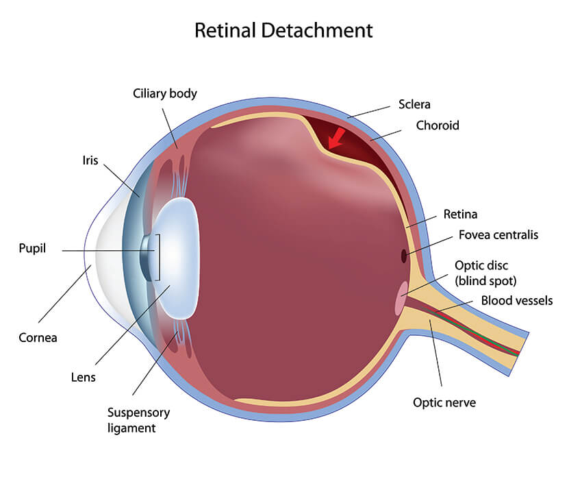
A retinal detachment occurs when a small hole or tear occurs in the retina and fluid from inside the eye travels through the tear and peels off the retina, detaching it from the eyewall.
A detachment can be partial or complete and needs immediate attention to minimize or avert vision loss. Treatment, usually done by a retina specialist, is often surgery to reattach the retina.
A retinal tear without a detachment can often be treated successfully with a laser. The laser will seal the tear and prevent a detachment. Symptoms of a retinal tear or detachment often start with flashes and floaters, as described below.

Flashes and Floaters
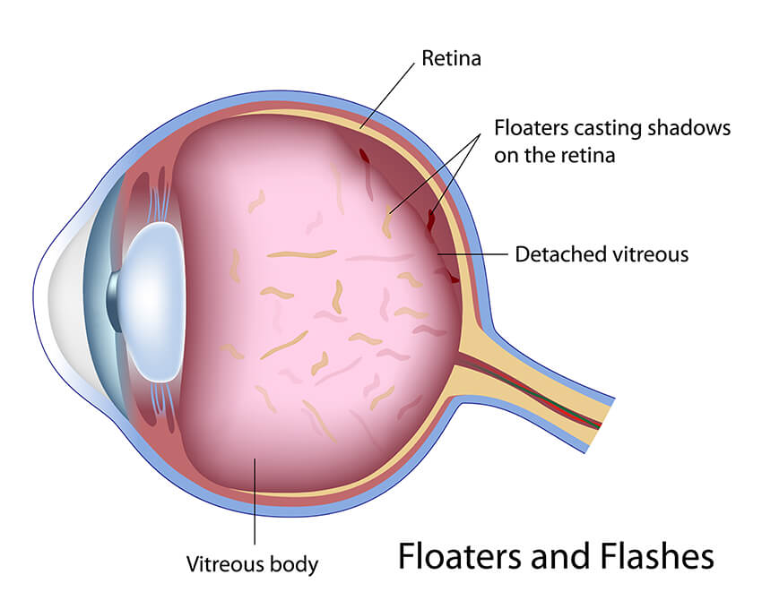
“Floaters” refer to debris or fibrils that are released by the vitreous when it pulls away from the retina. This “vitreous detachment” is a normal occurrence, and is often accompanied by flashes of light.
Floaters are commonly perceived by patients as seeing a “bug” or a “hair” in front of their eyes. If the detached vitreous pulls on the retina, the brain thinks the light is entering the eye, and flashes of light can be “seen.”
Although floaters and flashes are mostly innocent and do not represent a detached retina, a good retina examination is necessary to make sure a retinal tear has not occurred.

Macular Degeneration
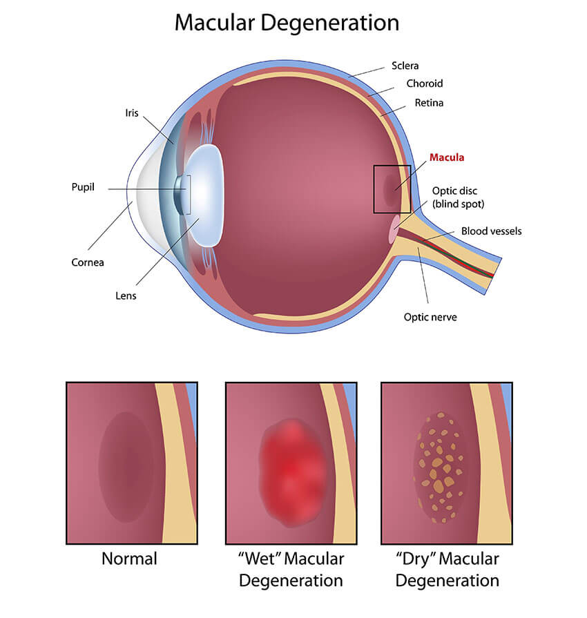
The macula is the center of the retina, similar to the bull’s eye being the center of a target. It is the most sensitive part of the retina and the part that gives precise vision for reading and driving. As we age, the macula can weaken and “degenerate,” causing vision impairment.
Macular degeneration can be divided into “wet” and “dry.” The cause of both types is not fully understood, but treatment is available in the form of AREDS 2 vitamins for the dry type, and laser or intraocular injections for the wet type.
Vision loss first occurs for reading and may necessitate strong reading glasses or magnifying aids. Modern diagnosis and treatment can now mostly prevent severe vision loss.


